


November 26, 2021
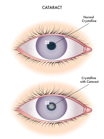
Any clouding or opacification of the natural crystalline lens in the eye is called cataract. There may be varying grades of opacification of the lens. Cataract surgery is indicated when the opacity, whatever grade it may be, interferes with the daily routine function of an individual.
A gradual loss of clear vision is the main symptom of a cataract. Vision for distance or near or for both may be blurred, objects may have fuzzy edges, lights at night may have coloured haloes. Sometimes, doubling of images may occur. Depending on the type of cataract, some of these changes are worse at night, some in daytime, and some all the time.
Increase in age is one of the most common cause for cataract. People with diabetes develop cataract at an early age compared to non-diabetic people. A number of other factors can influence cataract formation like injuries, inflammation in the eye, other diseases, use of some medications, and so on. Sometimes, cataract can be seen at or soon after birth and they are genetic in origin. There is unfortunately no proven treatment to prevent or slow the progression of age-related cataract.
When the daily routine functions are hampered due to the presence of cataract, it need to be removed surgically and replaced with Intra Ocular Lens (IOL) implantation. Maturity of cataract is no longer a criterion for cataract surgery due to technological advances and any grade of cataract can be removed when it interferes with functional vision.
Surgery is the only definitive treatment for cataract and cataract surgery can be done either by using ultrasound energy called Phacoemulsification or using femtosecond laser. The surgeon will remove the cataract through a very small opening in a painless manner. The small opening made closes on it own without any need for stitches. The operation can be done either using eye drops or an injection to anaesthetise the eye. A foldable lens (IOL) is inserted through the small opening. The new lens inside the eye allows a greatly improved quality of vision and minimises the need for glasses.
There are different types of IOLs like monofocal, multifocal, trifocal and Extended Depth of Focus IOL (EDF IOL) in the market. The choice of IOL depends upon the type of cataract, eye condition, cost and requirement of the patient.
Monofocal IOLs
These lenses provide good distance vision and most patients are not dependent on their distance glasses for daily activities. However, patients implanted with monodical IOLs typically require reading glasses after cataract surgery Toric IOLs These special lenses correct high cylindrical power reducing the patient’s dependence on distance glasses.
Multifocal IOLs
These lenses are designed to give good distance and near vision with less dependence on glasses.
Trifocal and Extended depth of focus IOLs
These type of IOLs give good intermediate vision which is necessary for many daily activities including computer usage, in addition to good distance and near vision.
No.However, in order to support the IOL inside the eye, a part of the natural lens called the posterior capsule is retained inside the eye. This membrane may sometimes increase in thickness or loses clarity many months to years after the cataract operation. When this happens, it can result in blurred vision. A simple OP procedure using laser clears the membrane and restore clear vision immediately.

Computer vision syndrome or digital eye strain refers to various symptoms related to prolonged use of computers , mobile phones etc. Nowadays most people work constantly with computers and it result in dry eyes, eye strain, tiredness, blurred vision, double vision, eye pain or headache. All or some of the symptoms can be present in a given individual and the symptom complex is called computer vision syndrome (CVS).
Characters in a computer screen are very bright at the center and diminish in intensity towards their edges. The edges are not well defined unlike printed material. Also the brightness is not uniform. This makes it very difficult for our eyes to remain focussed to the screen. When someone is focussing for a long time the eyes struggle to maintain focus and they tend to drift to a point called “resting point of accommodation”. This leads to the various symptoms of CVS.
While someone is working in the computer the blink rate is reduced. If the working place is air conditioned, it causes low humidity and this combined with long working hours with reduced blink rate leads to computer vision syndrome. Place the computer terminal below the level of your eyes, so that you look down at it. Looking up at the screen causes your upper lid to lift and exposes more of the ocular surface to the drying effects of a low humidity air-conditioned environment. Constant, conscious blinking when working on computers can help. Another useful thing to remember is the 20-20-20 rule. Every 20 minutes, one should look away from the screen into the distance (20 feet), for 20 seconds to rest the eye muscles.
Glare screen filters helps to reduce glare from the computer screen. An eye glass with anti reflective coating (AR) is also useful as the AR coating prevents glare and reflections. These may help somewhat, but they will not solve your computer vision problems completely because they only affect glare from the computer screen and not the visual problems related to the constant refocusing of your eyes when working at a computer.
A thorough eye examination is needed to confirm whether your symptoms are due to computer use alone. Examination includes vision assessment and the presence of any refractive error to decide the need to wear glasses for the distance at which the computer screen is viewed. Some may have reduced convergence ( the movement of eyes towards each other for near work) which needs to be treated by exercises. With increasing age, the tone of the muscle inside the eye that allows the natural lens to change shape is reduced. This leads to blurring of vision for near work which needs to be treated with glasses.The work environment and the design ergonomics of the desk and chair will also need to be determined as they can contribute to the symptoms mentioned. In addition to the above the eye lids and tear film status are to be assessed as lid margin disease or dry eyes can worsen the condition.
Make sure your sit in a comfortable chair with a good back rest. Room lighting should be free of glare and from the ceiling rather than from the side walls. Reduce very bright room lights so that the brightness and contrast of the computer screen can be reduced to comfortable levels. Working distance from eye to screen should be set between 50-60 cm. Viewing angle for the screen should be 10 degrees to 15 degrees below straight-ahead gaze position, to minimise the evaporation of tear film.
Any clouding or opacification of the natural crystalline lens in the eye isCornea is the clear, transparent structure in the front of the eye and along with Lens, it is responsible for focussing the light rays onto the nerve layer called Retina. Cornea is not a singe layer structure but it is a multilayered one. The transparency of the cornea can be affected due to various reasons. When the transparency and the resultant poor vision cannot be restored by medical treatment, glasses or contact lenses corneal transplantation is the surgical option to improve vision.
called cataract. There may be varying grades of opacification of the lens. Cataract surgery is indicated when the opacity, whatever grade it may be, interferes with the daily routine function of an individual.
A cornea transplant (keratoplasty) is a surgical procedure to replace part of the cornea of the patient with that of corneal tissue from a donor. A cornea transplant is most often used to restore vision to a person who has a damaged cornea from various causes. A cornea transplant may also relieve pain or other signs and symptoms associated with diseases of the cornea. Through eye donation program corneal tissue is removed from donors after death. The donor cornea tissue is thoroughly screened for any communicable diseases and examined for quality assessment. A cornea transplant can restore vision, reduce pain and improve the appearance of a damaged or diseased cornea.
With the advances in the field of Ophthalmology, it is now possible to remove only the affected layers of the cornea instead of replacing the entire, full thickness corneal tissue in selected patients. Avoiding a full thickness corneal transplantation reduces the complications and increase the survival rate of the graft. The various types of surgical procedures include
In this procedure the entire cornea from the patient is removed and replaced with the donor cornea. It is done for patients who have disease involving most or all the corneal layers.
This procedure removes diseased tissue from the back corneal layers called the Endothelium and Descemet membrane. In a healthy cornea, these layers help to keep the cornea dry and prevents fluid accumulation.In conditions where the disease process involves these deeper layers, thy can be selectively removed and replaced with the donor corneal layers.
There are two types of endothelial keratoplasty. The more common type, called Descemet Stripping Endothelial Keratoplasty (DSEK), uses donor tissue to replace about one-third of the cornea. A second type of procedure, called Descemet Membrane Endothelial Keratoplasty (DMEK), uses a much thinner layer of donor tissue.
There are different types of IOLs like monofocal, multifocal, trifocal and Extended Depth of Focus IOL (EDF IOL) in the market. The choice of IOL depends upon the type of cataract, eye condition, cost and requirement of the patient.
Monofocal IOLs
These lenses provide good distance vision and most patients are not dependent on their distance glasses for daily activities. However, patients implanted with monodical IOLs typically require reading glasses after cataract surgery Toric IOLs These special lenses correct high cylindrical power reducing the patient’s dependence on distance glasses.
Multifocal IOLs
These lenses are designed to give good distance and near vision with less dependence on glasses.
Trifocal and Extended depth of focus IOLs
These type of IOLs give good intermediate vision which is necessary for many daily activities including computer usage, in addition to good distance and near vision.
This procedure removes diseased tissue from the front corneal layers, namely epithelium and the stroma, but leaves the back endothelial layer in place. This is suitable for patients who are having corneal problems involving only the front portion of the cornea like a superficial scar. Depending upon the thickness and depth of the tissue removed, the procedure can be Superficial Anterior Lamellar Keratoplasty (SALK) or Deep Anterior Lamellar Keratoplasty (DALK). The removed tissue is replaced with the corresponding layers of donor cornea and secured in place with fine sutures. The back layers of the cornea are not disturbed and left intact.
Cornea transplant is a relatively safe procedure. Still, it does carry a small risk of serious complications, such as:
Cornea transplant is a relatively safe procedure. Still, it does carry a small risk of serious complications, such as:
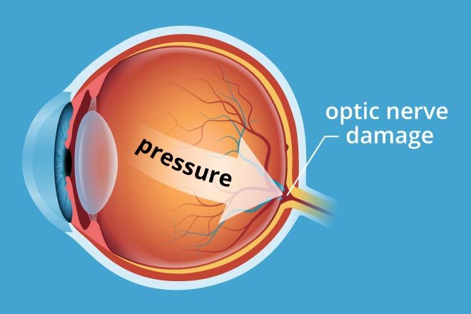
A complete pre op evaluation of both eyes is mandatory including visual acuity assessment, intra ocular pressure and examination under dilation to check the nerve fibre layer called retina and status of optic nerve. The risk factors are noted down and the consultant will discuss in detail about the surgical procedure, what to expect after the surgery, the post operative medications, frequency of follow up visits etc. Patients are advised to get one of the family members during the discussion to clarify any doubts regarding the surgical procedure. All the surgeries are done under local anaesthesia except in children where general anaesthesia is preferred. After baseline blood examination and assessment by the Anaesthesiologist, the patient is booked for the transplant procedure.
1.Control your blood sugar level
2.Maintain healthy blood pressure and cholesterol
3. Exercise regularly
4.Quit smoking
5. Dilated eye examination by your ophthalmologist at least once a year or more frequently depending upon the retinal changes present
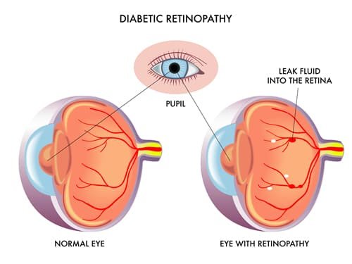
Maculopathy, threatening the centre of vision is treated with laser burns, which ‘dry up’ the waterlogged retina. Injections into the eye may also be helpful. Proliferative diabetic retinopathy is treated by the application of laser burns to the periphery of the retina. This reduces the volume of sick retina, whilst saving blood supply for the important central macular area. Bleeding into the vitreous of the eye may require vitrectomy, a surgical procedure in which the blood-stained gel is safely removed. Bleeding points are treated and laser applied to prevent the development of more abnormal ‘new vessels’. Modern vitrectomy surgery has revolutionised the treatment of severe proliferative diabetic retinopathy, giving hope to the most desperate of cases.
Poor vision for distant objects caused by refractive errors are the most common cause of visual impairment in children and often goes unnoticed by the parents. Symptoms of poor vision include squeezing eyes while watching TV or blackboard, holding things closely to see clearly, poor school performance, frequent complaints of headache and eye strain.
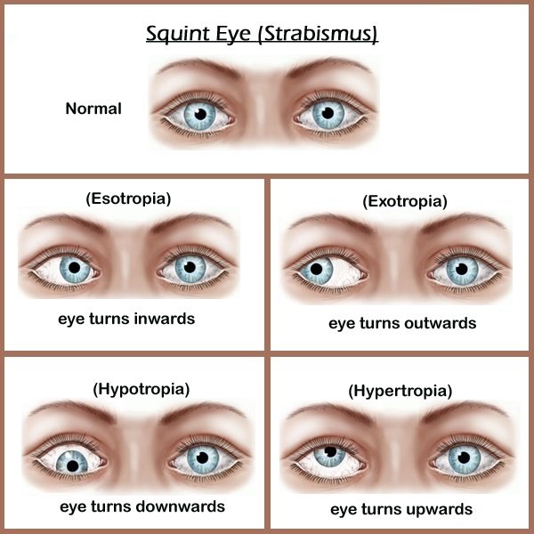
Deviation of eyes in which the eyes look in different directions instead of looking straight (Squint)
Watering with or without redness in one or both eyes
Drooping of eyelid( Ptosis)
Congenital cataract may be seen as a white reflex in the pupillary area and sometimes oscillating eye movements (Nystagmus)
Congenital glaucoma often presents with the child having watering of eyes, intolerance to light and large cornea
A dilated eye examination allows your Ophthalmologist to examine more thoroughly the retina and optic nerve for signs of damage before you notice any change to your vision.In the early stages the visual impairment may be mild but as the disease advances with more and more bleeding and swelling of the retina, it can lead to complications like intra ocular haemorrhage into vitreous cavity, retinal detachment and even permeant loss of vision.
Proliferative diabetic retinopathy is an advanced stage of the disease wherein inadequate oxygen supply to the retina results in the growth of abnormal blood vessels which bleed easily due to their fragile nature. This adds to the burden of already damaged retina and worsen the condition.
Modern screening methods will alert us to diabetic eye disease before the onset of symptoms. The decision to treat is made on the basis of clinical examination and special tests, including fluorescein angiography and OCT retinal scanning
Maculopathy, threatening the centre of vision is treated with laser burns, which ‘dry up’ the waterlogged retina. Injections into the eye may also be helpful. Proliferative diabetic retinopathy is treated by the application of laser burns to the periphery of the retina. This reduces the volume of sick retina, whilst saving blood supply for the important central macular area. Bleeding into the vitreous of the eye may require vitrectomy, a surgical procedure in which the blood-stained gel is safely removed. Bleeding points are treated and laser applied to prevent the development of more abnormal ‘new vessels’. Modern vitrectomy surgery has revolutionised the treatment of severe proliferative diabetic retinopathy, giving hope to the most desperate of cases.
1.Control your blood sugar level
2.Maintain healthy blood pressure and cholesterol
3. Exercise regularly
4.Quit smoking
5. Dilated eye examination by your ophthalmologist at least once a year or more frequently depending upon the retinal changes present
Maculopathy, threatening the centre of vision is treated with laser burns, which ‘dry up’ the waterlogged retina. Injections into the eye may also be helpful. Proliferative diabetic retinopathy is treated by the application of laser burns to the periphery of the retina. This reduces the volume of sick retina, whilst saving blood supply for the important central macular area. Bleeding into the vitreous of the eye may require vitrectomy, a surgical procedure in which the blood-stained gel is safely removed. Bleeding points are treated and laser applied to prevent the development of more abnormal ‘new vessels’. Modern vitrectomy surgery has revolutionised the treatment of severe proliferative diabetic retinopathy, giving hope to the most desperate of cases.
Home
About us
Our Treatments
Blogs
FAQ’s
Contact Us
Cataract
Cornea
Dry Eyes
Glaucoma
Refractive surgery
Specialty Contact lenses